How to Apply
Applications are closed for 2017. Interested in taking the course in the future? Sign up for our mailing list (at bottom of page) to be notified when we start accepting applications for next year.
Course Description
The Zeiss Berkeley Brain Microscopy Innovation Center at the University of California, Berkeley offers an annual course on state-of-the-art optical tools, imaging systems, and analysis methods that are being developed for the study of neuronal circuits. Our intensive week-long course includes training in the theory and practice of emerging BRAIN Initiative neurotechnologies. Up to 16 students will work side by side with Berkeley Neuroscience faculty as they rotate through microscope demos and then choose a microscope station for a project.
Agenda
We will post the 2017 agenda at the conclusion of the course. For now, you can look at the following to get a feel for what the course is like.
>Download a pdf of last year’s agenda.
Microscope Demos
Project Rotations
Students will rotate through demonstrations of eight microscope stations before choosing one station for a three day project. The eight microscope stations and demos are:
Population Calcium Imaging in Retina
Microscope: Two-photon scanning microscope with DMD for visual stimulation.
Demo: Two-photon calcium imaging of light responses in retina.
Station Leaders: Marla Feller, Ben Smith, Franklin Caval-Holme
Two-Photon Optogenetics With Holography
Microscope: 3i in vivo 2P Microscope (2P Scanning Imaging with 2P Temporal Focusing Holography, AKA Millennium Falcon).
Demo: Two-photon excitation of neurons using temporally focused holography and soma-targeted opsins.
Station Leaders: Ben Shababo
CUBIC Tissue Clearing Demo
Microscope: Fluorescence Light Sheet Microscopy.
Demo: The CUBIC protocol will be used to clear whole mouse embryos or whole mouse brains. Fluorescence light sheet microscopy follows after processing.
Station Leaders: Eva Nichols
Light Sheet Fluorescence Microscopy
Microscope: Zeiss LightSheet.Z1 Selective Plane Illumination Microscopy (AKA Morpheus).
Demo: Basic introduction to differences and benefits of light sheet microscopy vs. epi-illumination. Sample handling and positioning large 3D specimens such as cleared brain tissue. Acquiring z-stacks and 3D-rendering large datasets.
Station Leaders: Scott Olenych
Voltage Imaging of Neuronal Activity
Microscope: Zeiss Axio Observer inverted widefield microscope equipped with a Hammamatsu sCMOS camera. For simultaneous, multicolor imaging we use a Photometrics dual-view emission splitter. Hardware is controlled via Micro-Manager software.
Demo: In this exercise, we will primarily use voltage-sensitive dyes, synthesized in the Miller Lab, combined with widefield microscopy which has been optimized for the fast and sensitive recording of electrical signals. Utilizing dissociated hippocampal neurons, we will record evoked and spontaneous action potential firing, interface voltage imaging with complimentary fluorescent tools and discuss the technical limitations of this method. We will also demonstrate image processing and spike detection analyses using Matlab, a key element to the application voltage imaging techniques.
Station Leaders: Alison Walker, Rishi Kulkarni
Imaging Embryonic Zebrafish Spinal Cord Activity
Microscope: Zeiss LSM 880 (AKA Neo), Coherent Chameleon 2p laser.
Demo: In vivo imaging the spinal cord of a one-day-old zebrafish expressing GCaMP and other fluorescent indicators.
Station Leaders: Amy Winans, Jen Lee
Optical Quantal Analysis of Synaptic Function and Plasticity
Microscope: Spinning Disk Confocal with 1p spatial light modulator (SLM) Holography (AKA Enterprise) and Zeiss Axiozoom Wide Field Microscope for in vivo calcium imaging.
Demo: In this demo we will use use a combination of microscopes to demonstrate how to measure synaptic activity and plasticity with both single synaptic vesicle and single synapse resolution in an intact Drosophila larva. Additionally we will demonstrate how these imaging methods can be coupled with 1P SLM holography photo-activation and photo-uncaging.
Station Leaders: Zachary Newman, Adam Hoagland
Fast Confocal Imaging with Zeiss Fast Airyscan
Microscope: Inverted LSM 880 with Airyscan and Fast Airyscan.
Demo: This station will demonstrate the latest Airyscan (superresolution with confocal) and Fast Airyscan technologies compared to standard confocal.
Station Leaders: Renee Dalrymple
Survey Demos
Students will also rotate through short demos on the following seven microscope stations to get a survey of additional approaches for imaging and manipulating brain circuits:
Imaging Transcriptional Dynamics in Fly Embryos
Microscope: Leica SP8 confocal
Demo: We’ll be using a confocal microscope to image transcription of important developmental genes in a living fly embryo.
Station Leaders: Armando Reimer, Yang Joon Kim
3D Holographic Temporal Focusing
Microscope: Two-photon spatial light modulator holography
Demo: We will show how to photo-stimulate individual neurons with 3D-SHOT, a two-photon holography method with temporal focusing.
Station Leaders: Nicolas Pegard
Computational Microscopy using LED Array Illumination
Microscope: Nikon Inverted Microscope, PCO.edge 5.5 SCMOS Camera, Custom LED Array (Quasi-Dome).
Demo: We will demo our new prototype LED Array microscope, a modification of a conventional LED Array microscope where the lamp of a regular microscope is replaced with an LED array, allowing many new capabilities. We will show brightfield, darkfield, and phase contrast imaging all using computational illumination which can be changed quickly with minimal hardware modifications. We will also show results for super-resolution imaging using Fourier Ptychography, which enables 5x better resolution by “cheating” the diffraction limit of the microscope using the same illumination configuration.
Station Leaders: Zack Phillips, Michael Chen
Imaging Near-Infrared Nanosensors for Dopamine Detection in Brain Tissue
Microscope: We will be using an inverted Zeiss Axio-Observer, Princeton Instruments NIRvana 2d InGaAs camera array, a Princeton Instruments IsoPlane spectrograph and Princeton Instruments PyLon 1d InGaAs array detector.
Demo: Nanosensors comprised of functionalized single-walled carbon nanotubes (SWNT) are a new class of near-infrared fluorophores capable of detecting the neurotransmitter dopamine. In this module, we will demonstrate the use of a custom built, near-infrared microscopy and spectroscopy suite for imaging and detecting changes in dopamine concentration in highly-scattering tissue phantoms using SWNT nanosensors.
Station Leaders: Jackson Travis Del Bonis-O’Donnell, Abraham Beyene
In Vivo Calcium Imaging with Optogenetic Stimulation
Microscope: 3i VIVO Spinning disk confocal with Phasor computer-generated holography (AKA Enterprise).
Demo: We will combine GCaMP imaging in live larval zebrafish with optogenetic stimulation using computer-generated holography to stimulate multiple cells simultaneously.
Station Leaders: Alden Conner
Fluorescence Lifetime Imaging of Small Molecules and Proteins
Microscope: Laser scanning confocal (LSM 510) capable of both one- and two- photon imaging, as well as fluorescence lifetime measurements (AKA Deckard).
Demo: Fluorescence lifetime imaging (FLIM) to measure absolute membrane potential will be demonstrated using small molecule voltage-sensitive dyes. FLIM will be used to record the voltage response to growth factor signaling in living cells.
Station Leaders: Julia Lazzari-Dean
Spectral Imaging & Linear Unmixing
Microscope: LSM 780 Confocal and Linear Unmixing within the Zen software package.
Demo: We will collect full spectral data from fluorescence samples and then show how this data can be computationally analyzed to quickly and efficiently separate fluorescent signals, even when the spectra have extensive overlap.
Station Leaders: Nipam Patel
Lecture Topics and Speakers
Lectures will cover the theory and research methodology underlying the development and use of imaging technologies for the study of neural circuits.
- Introduction to Optics for Microscopy, Paul Herzmark
- Microscopy Methods for Neurophysiology, Marla Feller
- Neural Circuits, Dan Feldman
- Photochemical and Optogenetic Tools for Measuring and Manipulating Neural Activity, Ehud Isacoff
- Optical Quantal Analysis, Zachary Newman
- Optical Dissection of Astrocyte-Neuron Circuits, Kira Poskanzer
- Circuit Mapping, Hillel Adesnik
- Building Your Witty Lab: Tools for Analyzing Advanced Microscopy of Brain, Dani Ushizima
- Tutorial on Deep Learning and Image Segmentation, Maryana Alegro
- Linear Unmixing, Nipam Patel
- Cameras and Detectors, Ben Smith
- Modeling Epilepsy and Imaging Seizures in Zebrafish, Scott Baraban
- Computational Microscopy for Neural Activity Tracking, Laura Waller
- FRET, Holly Aaron
- Fluorophore and BioSensor Design, Evan Miller
- Fluorescent Lifetime Imaging (FLIM), Julia Lazarri-Dean
- Clearing Agents & Techniques, Eva Nichols
- Introduction to Image Processing, Lynsey Hamilton
- Adaptive Optics, Na Ji
- Super Resolution Techniques, Xavier Darzacq
- Non-linear Optical Microscopy for Quantitative Cell Imaging, Aaron Streets
- Data Visualization & Presentation, Jen-Yi Lee
- Keynote Talk, David Schaffer
Faculty
The following are confirmed faculty for 2017. Check back for updates.
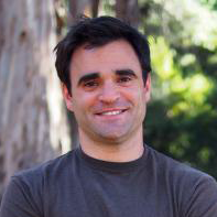 | Hillel Adesnik | Assistant Professor of Neurobiology | Research Focus: Neural basis of sensation, perception, and action. |
 | Scott Baraban | Professor, Department of Neurological Surgery at UCSF | Research Focus: Understanding the fundamental basis of epilepsies. |
 | Xavier Darzacq | Assistant Professor of Genetics, Genomics and Development | Research Focus: Mechanisms of transcription regulation. |
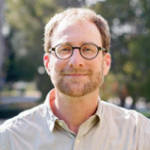 | Dan Feldman | Professor of Neurobiology | Research Focus: Sensory processing and plasticity in the somatosensory cortex. |
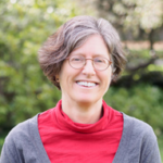 | Marla Feller | Professor of Neurobiology | Research Focus: Functional development and organization of neural circuits in the retina. |
 | Ehud Isacoff | Professor of Neurobiology | Research Focus: Mechanisms of ion channel function, synapse development, plasticity, and neural circuit function. |
 | Na Ji | Associate Professor of Physics and Molecular and Cell Biology | Research focus: Imaging technology such as adaptive optics for the study of neural circuit activity. |
 | Evan Miller | Assistant Professor of Chemistry and Molecular and Cell Biology | Research Focus: Development and application of molecular tools for studying neuroscience. |
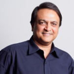 | Nipam Patel | Professor of Genetics, Genomics and Development | Research Focus: Developmental basis of evolutionary change. |
 | Kira Poskanzer | Assistant Professor of Biochemistry and Biophysics at UCSF | Research Focus: Astrocytic and neuronal function in the cortical microcircuit. |
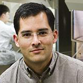 | David Schaffer | Professor of Chemical and Biomolecular Engineering, Bioengineering | Research Focus: Engineering stem cell and gene therapeutics. |
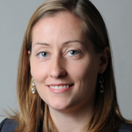 | Laura Waller | Associate Professor of Electrical Engineering and Computer Sciences | Research Focus: designing imaging systems and algorithms. |
Registration Information
If you are chosen to participate in the course, we will contact you with information on how to register, as well as information on travel and housing. A registration fee of $1200 covers the cost of the course and all meals.
Contact and Mailing List
The course organizers are Holly Aaron (Director of the BrainMIC and Molecular Imaging Center), Jen-Yi Lee (Molecular Imaging Center), and Georgeann Sack (Berkeley Neuroscience Communications). You can reach us by email at brainmic@berkeley.edu.
Interested in receiving updates about the course now and in future years? Join our mailing list. You can unsubscribe at any time.

